Home
Uncategories
Cross Section Of A Bone / Researchers Describe Mechanism That Underlies Age Associated Bone Loss Medica World Forum For Medicine : The upper (biting) surfaces of the tooth are at top, with the lower sections (bottom) embedded in the gums and jaw bone (not shown).
Cross Section Of A Bone / Researchers Describe Mechanism That Underlies Age Associated Bone Loss Medica World Forum For Medicine : The upper (biting) surfaces of the tooth are at top, with the lower sections (bottom) embedded in the gums and jaw bone (not shown).
Cross Section Of A Bone / Researchers Describe Mechanism That Underlies Age Associated Bone Loss Medica World Forum For Medicine : The upper (biting) surfaces of the tooth are at top, with the lower sections (bottom) embedded in the gums and jaw bone (not shown).. Select from premium bone cross section of the highest quality. And why does the marrow stop where it does, and so sharply? The central tubular region of the bone, called the diaphysis. The large dark spots are passages for blood vessels and nerves. In addition, cortical bone thickness at anterior, posterior, medial, and lateral parts of the bone section was measured.
Bone matrix and cells bone matrix osseous tissue is a connective tissue and like all connective tissues contains relatively few cells and large amounts of extracellular matrix. Now that you know what bones do, let's take a look at what they're made of and their anatomy. There are three general classes of bone. Bone and bones / pathology*. To the left is muscle tissue, and to the right is bone marrow.
Bone Structure Anatomy And Physiology I from s3-us-west-2.amazonaws.com Bone on side of the foot Bone markings the surface features of bones vary considerably, depending on the function and location in the body. Bone structure lower back 12 photos of the bone structure lower back back bone structure lower back, bone structure lower back, human bone structure lower back, lower back and hip bone structure, skeletal structure lower back, bone, back bone structure lower. Histology slide courtesy of william l. Related posts of cross section of a long bone foot bone anatomy x ray. I don't find it enhances the image. The upper (biting) surfaces of the tooth are at top, with the lower sections (bottom) embedded in the gums and jaw bone (not shown). This is essentially a measure of how the material is distributed about a given axis.
Bone cross section illustrations & vectors.
In the center of each osteon is the central canal, a space that houses blood vessels and nerves that supply bone. While it is not as hard as compact bone, spongy bone plays an important role of protecting the marrow where blood cells are produced. The geometrical properties generated from the ct image included as follows: This is essentially a measure of how the material is distributed about a given axis. The compact bone is made up of osteon. Decalcified compact bone looks completely different than compact bone that still has calcium salts in its matrix. Bone on side of the foot Browse 4,291 bone cross section stock photos and images available, or search for human bone cross section to find more great stock photos and pictures. Compact bone, spongy bone, and bone marrow. There are trabeculae in spongy bone which gives its sponge like appearance. Marrow in the shaft of long bones is typically yellow, with red marrow in the head through the cancellous bone. I don't find it enhances the image. As the names suggest compact bone looks compact and the spongy bone looks like sponges.
The geometrical properties generated from the ct image included as follows: Histology slide courtesy of william l. After a fracture, woven bone forms initially and is gradually replaced by lamellar bone during a process known as bony substitution. A cross section of a human long bone. Now that you know what bones do, let's take a look at what they're made of and their anatomy.
A Femur Cross Section Anatomy Of The Femur Cross Section Anatomy Medicine Com from anatomy-medicine.com The skeleton consists of bones connected at joints, or articulations, and is subdivided into two divisions. The upper (biting) surfaces of the tooth are at top, with the lower sections (bottom) embedded in the gums and jaw bone (not shown). The large dark spots are passages for blood vessels and nerves. Histology slide courtesy of william l. And why does the marrow stop where it does, and so sharply? As the names suggest compact bone looks compact and the spongy bone looks like sponges. Decalcified compact bone looks completely different than compact bone that still has calcium salts in its matrix. Related posts of cross section of human bone diagram bone structure lower back.
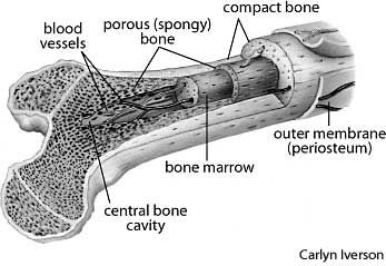
A diagrammatic view of a cross section of bone.
Compact bone is the outer layer and the spongy bone forms the inner layer. Bone and bones / pathology*. Each bone in your body is made up of three main types of bone material: Decalcified compact bone looks completely different than compact bone that still has calcium salts in its matrix. Section of bone marrow affected by myeloma seen under a microscope. The cross section of this circular cylinder is a circle. Bone structure lower back 12 photos of the bone structure lower back back bone structure lower back, bone structure lower back, human bone structure lower back, lower back and hip bone structure, skeletal structure lower back, bone, back bone structure lower. This is known as the periosteum. I = ∫ (y2 δa). Related posts of cross section of human bone diagram bone structure lower back. Learn vocabulary, terms, and more with flashcards, games, and other study tools. Learn vocabulary, terms, and more with flashcards, games, and other study tools. There are three general classes of bone.
Section of bone marrow affected by myeloma seen under a microscope. The surface features of bones vary considerably, depending on the function and location in the body. Bone markings the surface features of bones vary considerably, depending on the function and location in the body. Cross section of mandible at first molar region showing cortical and spongy bone basic concepts in osteogenesis. I don't find it enhances the image.
Schematic Diagram Of Long Bone Cross Section 47 Download Scientific Diagram from www.researchgate.net This is known as the periosteum. Compact bone is the outer layer and the spongy bone forms the inner layer. The central tubular region of the bone, called the diaphysis. I don't find it enhances the image. As the names suggest compact bone looks compact and the spongy bone looks like sponges. Bone markings the surface features of bones vary considerably, depending on the function and location in the body. To the left is muscle tissue, and to the right is bone marrow. While it is not as hard as compact bone, spongy bone plays an important role of protecting the marrow where blood cells are produced.
There are trabeculae in spongy bone which gives its sponge like appearance.
In the center of each osteon is the central canal, a space that houses blood vessels and nerves that supply bone. Section of bone marrow affected by myeloma seen under a microscope. This is essentially a measure of how the material is distributed about a given axis. Start studying cross section of long bone. Body size standardization was done, using the following equations: Smartdraw includes 1000s of professional healthcare and anatomy chart templates that you can modify and make your own. This slide contained a cross section of a very small bone, and you are looking at the entire thickness of the shaft of the bone. Haz tu selección entre imágenes premium sobre bone cross section de la más alta calidad. I don't find it enhances the image. Histology slide courtesy of william l. They are obtained by taking imaginary slices perpendicular to the main axis of organs, vessels, nerves, bones, soft tissue, or even the entire human body. The geometrical properties generated from the ct image included as follows: The two ends of the diaphysis are involved in forming joints.


0 Comments:
Posting Komentar